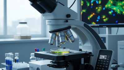
2026 Top Live Cell Imaging Microscope Types

In 2026, live cell imaging microscope systems are becoming more specialized, faster, and easier to compare. Yet the best choice still depends on the biological question. Researchers may select widefield microscopy for rapid screening, confocal microscopy for optical sectioning, or spinning-disk systems for gentler long-term observation. TIRF microscopy can reveal membrane events near the coverslip. Light-sheet platforms reduce phototoxic exposure in larger specimens. Super-resolution systems uncover molecular details that conventional optics cannot resolve.

Eric Betzig, a Nobel Prize-winning physicist and microscopy pioneer, described the purpose of advanced imaging clearly: “The goal of microscopy is to see more clearly, and to see more deeply.” His statement remains practical for modern live-cell research. Better resolution alone does not guarantee better data. Photobleaching, temperature drift, fluorescent labeling, and cell movement can distort an attractive image. Small errors matter.

This guide compares the major live cell imaging microscope types expected to shape laboratory workflows in 2026. It considers resolution, imaging speed, phototoxicity, sample compatibility, automation, and total operating cost. These factors often conflict. A high-resolution system may stress delicate cells. A fast platform may sacrifice structural detail. No single instrument wins every experiment.

That is the difficult part.

Reliable selection requires more than reading specifications. It requires matching microscope performance with cell behavior, experimental duration, and the evidence needed for publication. Some comparisons remain imperfect because manufacturers report performance under different conditions. Readers should verify results with application data, independent testing, and experienced microscopy specialists.
2026 Top Live Cell Imaging Microscope Types

Definition and Role of Live Cell Imaging Microscopes

2026 Top Live Cell Imaging Microscope Types

Definition and Role of Live Cell Imaging Microscopes

Live cell imaging microscopes observe living cells without fixing or staining them permanently. They record movement, division, transport, and shape changes over time. Their role is not merely visual. These systems connect cellular structure with biological function under controlled conditions.

Common types include phase-contrast, widefield fluorescence, confocal, spinning-disk, TIRF, and light-sheet microscopes. Phase contrast suits routine monitoring with low phototoxicity. Confocal systems provide optical sectioning but may stress sensitive cells during long experiments. Spinning-disk designs often balance speed and reduced light exposure. Light-sheet systems can limit illumination, although sample preparation remains demanding.

Industry data reflects rising demand. A 2024 MarketsandMarkets report estimated the live-cell imaging market would grow strongly through 2029, supported by drug discovery and cell biology research. Grand View Research also reported continued market expansion, driven by automated imaging and advanced fluorescence methods. These figures are useful, but forecasts are not laboratory evidence. They can overlook maintenance costs, calibration time, and operator skill.

Practical Considerations

In practice, microscope selection depends on the question. A 24-hour migration assay may need gentle phase contrast. Fast vesicle transport may require high-speed fluorescence. Thick organoids often benefit from optical sectioning or light-sheet illumination. Better resolution is not always better biology. I have seen beautiful images fail when cells were overheated, overexposed, or poorly controlled. Temperature, carbon dioxide, illumination dose, and imaging intervals deserve equal attention.

Core Optical Principles Used in Live Cell Observation

2026 Top Live Cell Imaging Microscope Types

Core Optical Principles Used in Live Cell Observation

Live cell imaging begins with managing light, contrast, and specimen stress. Brightfield microscopy uses transmitted light to reveal cell outlines, but transparent structures may remain faint. Phase contrast converts small phase shifts into visible intensity differences. It works well for observing cell shape, attachment, and movement without fluorescent labels.

Differential interference contrast adds directional contrast and creates a relief-like image. The effect is useful for monitoring edges, vesicles, and dividing cells. Fluorescence microscopy uses excitation and emission filters to isolate labeled molecules. Widefield illumination is fast, but light above and below the focal plane can reduce clarity. Optical sectioning improves this issue. Confocal systems reject much out-of-focus light through a pinhole, producing sharper sections from thicker samples.

Total internal reflection fluorescence restricts excitation near the coverslip. It is valuable for studying membrane events and early cell attachment. Light-sheet microscopy illuminates a thin plane from the side, reducing exposure during long observations. Phototoxicity still matters. Light dose matters.

From practical imaging work, stable temperature, humidity, and focus often influence results more than higher resolution. A microscope may show impressive detail while gradually harming the cells. I still find focus drift easy to underestimate, especially during overnight experiments. Controls should include unlabeled cells, illumination tests, and repeated fields of view. Optical performance is only reliable when cell health, acquisition settings, and analysis methods are checked together.

Major Types of Live Cell Imaging Microscopes in 2026

Major Types of Live Cell Imaging Microscopes in 2026

Live cell imaging microscopes now serve different biological questions. Widefield fluorescence is fast and gentle, making it useful for membrane movement and long time-lapse studies. Phase-contrast and DIC systems avoid fluorescent labels, so they suit cell growth and morphology checks. Confocal microscopy provides optical sections, but repeated laser exposure can stress sensitive cells. Spinning-disk confocal reduces exposure and captures faster cellular events.

Light-sheet microscopy limits illumination outside the focal plane. This design supports longer observations of organoids, embryos, and three-dimensional cultures. TIRF microscopy focuses near the coverslip, revealing vesicle docking and cytoskeletal activity.

According to Grand View Research, the global live cell imaging market was valued at about USD 2.3 billion in 2023 and is projected to grow at roughly 8.7% annually through 2030. MarketsandMarkets also identifies fluorescence, high-content screening, and three-dimensional imaging as major growth areas.

Tips: Match the microscope to the biology, not the resolution chart. Test phototoxicity with a short pilot run. Record temperature, carbon dioxide, exposure time, and medium composition. No platform wins every experiment. A highly sensitive camera cannot repair poor focus or unstable incubation. I still find this easy to underestimate. In practice, the quietest system often produces the most trustworthy movie.

How to Compare Resolution, Speed, and Cellular Compatibility

2026 Top Live Cell Imaging Microscope Types

Comparing live-cell microscopes starts with the biological question, not the sharpest image. Widefield systems can capture fast events with low light exposure, but background rises in thick samples. Spinning-disk confocal imaging improves optical sectioning and often records rapid cellular dynamics with less phototoxicity than point scanning. Light-sheet microscopy reduces illumination outside the focal plane, making it valuable for long 3D observations. TIRF excels near cell membranes, while multiphoton systems reach deeper tissue with infrared excitation. The choice is not perfectly clean.

Resolution depends on numerical aperture, wavelength, sampling, and specimen movement. Conventional fluorescence commonly provides about 200 nanometers of lateral resolution, while live-cell super-resolution may reach roughly 20–100 nanometers under controlled conditions (Sahl et al., Science, 2017). Speed also needs a practical measure. A 10-frame-per-second system is useless if cells bleach after two minutes. The 2024 Grand View Research microscopy market analysis valued the sector at approximately 8 billion US dollars, with live-cell and high-content applications among key growth areas. MarketsandMarkets also identifies automated cellular imaging as a major expansion segment in its 2024 analysis. These figures describe demand, not experimental quality. Cellular compatibility remains decisive: check temperature stability, carbon dioxide control, mechanical drift, illumination dose, and medium exchange. A beautiful image can still be biologically wrong. For fragile cells, lower light and longer observation may outperform nominal resolution.

2026 Top Live Cell Imaging Microscope Types - How to Compare Resolution, Speed, and Cellular Compatibility

Representative performance ranges for commonly used live-cell microscopy approaches. Actual results depend on numerical aperture, fluorophore, detector sensitivity, sample thickness, environmental control, and acquisition settings.

Microscope Type Typical Lateral Resolution Typical Axial Resolution Practical Imaging Speed Optical Sectioning Phototoxicity and Photobleaching Cellular Compatibility Best-Fit Applications
Widefield Fluorescence Approximately 200–300 nm Approximately 500–1,000 nm; out-of-focus fluorescence is collected Very high; commonly 10–100+ frames per second for small regions, depending on signal and camera Low without computational deconvolution Low to moderate
Efficient illumination can reduce exposure dose, but the entire field and sample depth are illuminated.
Excellent for adherent cells, labeled organelles, and low-light-sensitive samples; compatible with standard culture chambers Fast 2D dynamics, cell motility, mitosis, membrane movement, and long-term fluorescence imaging
Spinning-Disk Confocal Approximately 180–250 nm Approximately 500–800 nm High; often 10–50 frames per second for small fields, with rapid optical sectioning Good; multiple pinholes reject much of the out-of-focus signal Low to moderate
Usually lower than point-scanning confocal because illumination is distributed across many pinholes.
Very good for living cells, organelles, and moderately thick specimens; requires sufficient fluorescence signal Organelle trafficking, vesicle transport, cytoskeletal dynamics, and 3D imaging of cell monolayers
Laser-Scanning Confocal Approximately 180–250 nm Approximately 500–800 nm Moderate; commonly 0.5–10 frames per second for full-frame imaging, depending on scan mode and resolution Excellent; optical section thickness can be adjusted with the pinhole Moderate to high
Point illumination can deliver a relatively high local dose, especially during rapid or repeated z-stacks.
Good for robust fluorescent labeling and thicker samples; careful dose management is important for long-term experiments High-contrast 3D imaging, z-stacks, subcellular localization, and moderately thick tissues or organoids
TIRF Microscopy Approximately 150–250 nm in the lateral plane Approximately 70–200 nm effective excitation depth Very high; frequently 10–100 frames per second for suitable fields and signals Excellent near the coverslip; excitation is restricted to a shallow evanescent field Low to moderate
Low background and shallow excitation reduce exposure outside the imaging plane.
Excellent for cells attached to a clean coverslip; limited for structures deeper than roughly 100–200 nm from the interface Exocytosis, endocytosis, focal adhesions, membrane signaling, and single-particle or single-molecule studies near the basal membrane
Light-Sheet Fluorescence Approximately 200–400 nm, depending on illumination geometry and objective Approximately 500–1,500 nm, depending on the light-sheet thickness and detection optics High for volumetric imaging; tens to hundreds of planes per second are possible in specialized configurations Excellent; illumination is confined to the plane being observed Low per volume
Reduced out-of-plane illumination is advantageous for long-term imaging, although alignment and sample handling are more demanding.
Excellent for embryos, organoids, cleared or lightly scattering specimens, and larger 3D samples; conventional adherent-cell workflows may require special mounting Long-term 3D development, organoid morphology, cell migration, and volumetric imaging of delicate specimens
Structured Illumination Microscopy Approximately 100–150 nm laterally in conventional 2D SIM Approximately 250–400 nm axially in 3D SIM Moderate to high; commonly 1–20 reconstructed frames per second, depending on the number of raw images and field size Good to excellent; optical sectioning is combined with computational reconstruction Moderate
Lower than many point-scanning methods, but multiple raw exposures are required for each reconstructed image.
Good for fluorescently labeled live cells with adequate signal and limited movement during acquisition; reconstruction artifacts can occur with rapid motion Near-diffraction-limited dynamics, cytoskeletal organization, organelle contacts, and fine cellular structures
Phase-Contrast or DIC Approximately 250–350 nm for visible cellular features, limited by contrast rather than fluorescence specificity Limited optical sectioning; typically suited to thin specimens Very high; video-rate imaging is readily achievable with modern cameras Low; DIC provides directional contrast rather than true optical sectioning Very low
No fluorescent label or excitation light is required.
Excellent for unlabeled cells, primary cultures, and long-term monitoring; compatible with standard incubated culture vessels Cell growth, morphology, migration, division, confluence, and label-free viability monitoring
Two-Photon Fluorescence Approximately 300–500 nm, depending on wavelength and numerical aperture Approximately 700–1,500 nm Low to moderate; often 0.1–5 frames per second for full-frame images, with faster targeted acquisition possible Excellent; excitation is intrinsically localized to the focal region Low to moderate in depth
Reduced out-of-focus excitation benefits thick samples, but high peak intensities can damage sensitive specimens.
Very good for thick, scattering, or three-dimensional living samples, including tissue slices and organoids; requires suitable fluorophores Deep-tissue imaging, neuronal activity, organoid structure, and long-term observation below the sample surface

Resolution values are representative ranges for visible-light systems using appropriate high-quality objectives and properly sampled images. They should be treated as comparison guidelines rather than guaranteed specifications.

Choosing a Microscope for Different Live Cell Imaging Tasks

Choosing a live-cell microscope starts with the biological question, not the camera. For rapid migration, widefield fluorescence offers high frame rates and simpler illumination. It can reduce phototoxicity, although out-of-focus glow may weaken measurements. Confocal imaging suits thick samples needing optical sections. Spinning-disk systems are often gentler for fast 3D imaging.

For membrane trafficking, total internal reflection fluorescence isolates events near the coverslip, sometimes within roughly 100–200 nanometers. Light-sheet microscopy is better for organoids and embryos because it illuminates only the observed plane. According to Grand View Research’s 2024 market analysis, the global live-cell imaging market was valued at more than USD 2 billion in 2023 and is projected to grow strongly through 2030. That growth reflects demand for faster, less damaging measurements, not merely sharper pictures.

Practical testing matters. Record a five-minute time-lapse before committing to an experiment. Check focus drift, temperature stability, stage vibration, and cell survival after illumination. Use environmental control for long experiments. A 2024 MarketsandMarkets report identified three-dimensional imaging and automated analysis as major growth areas in microscopy. Yet automation cannot repair poor contrast or dying cells. I would also question impressive resolution claims when the sample moves, bleaches, or changes shape. Sometimes, a modest widefield setup produces more trustworthy data than a complex system used without careful controls.

2026 Top Live Cell Imaging Microscope Types

Choosing a Microscope for Different Live Cell Imaging Tasks

Scores use a 1–5 comparative suitability scale based on established modality characteristics. Widefield and spinning-disk confocal systems are commonly suited to fast routine imaging, TIRF is optimized for near-membrane events, light-sheet microscopy is designed for long-duration 3D imaging with reduced phototoxicity, and multiphoton microscopy is advantageous for thicker specimens.

Search engine powered by ElasticSuite