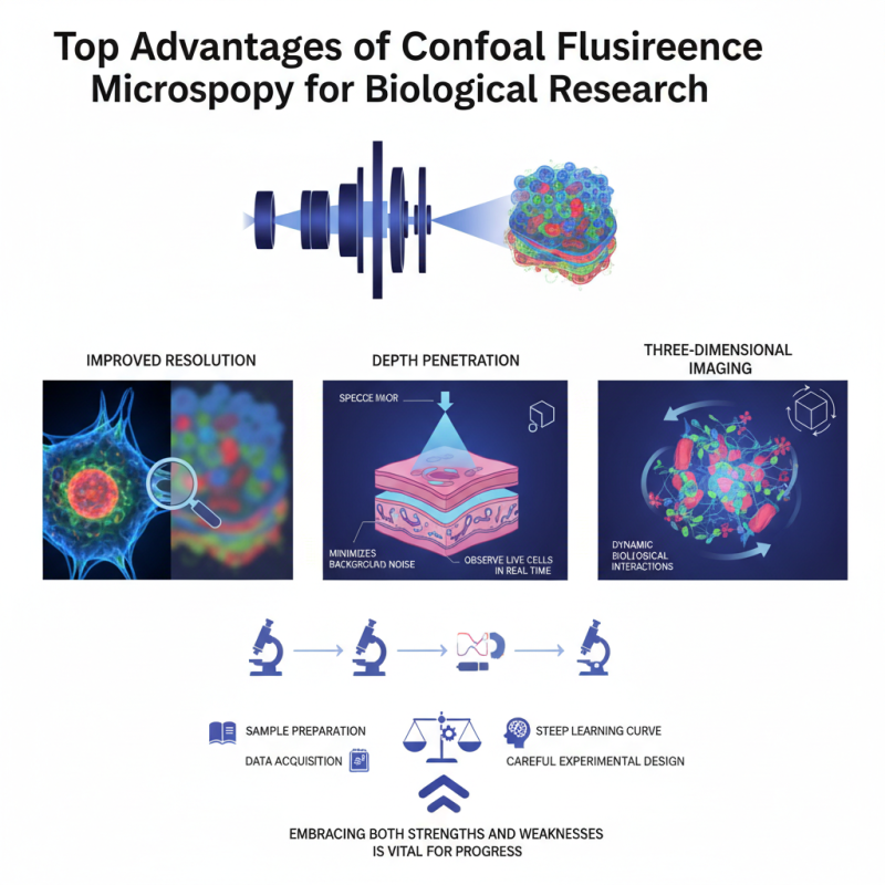
Confocal fluorescence microscopy has revolutionized biological research in recent years. This powerful technique allows scientists to observe specimens in greater detail. By utilizing focused laser beams, researchers capture high-resolution images. The ability to visualize specific structures enhances understanding of cellular processes.
This method excels in three key areas: improved resolution, depth penetration, and three-dimensional imaging. Confocal fluorescence microscopy minimizes background noise, resulting in clearer images. Researchers can observe live cells in real time, providing insights into dynamic biological interactions. However, the technique has limitations that can hinder certain studies.
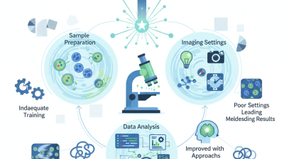
Despite its advantages, confocal fluorescence microscopy requires careful experimental design. It demands a steep learning curve for proper execution. Researchers must always check for potential pitfalls. Balancing sample preparation and data acquisition is crucial. As the field evolves, embracing both strengths and weaknesses is vital for progress.
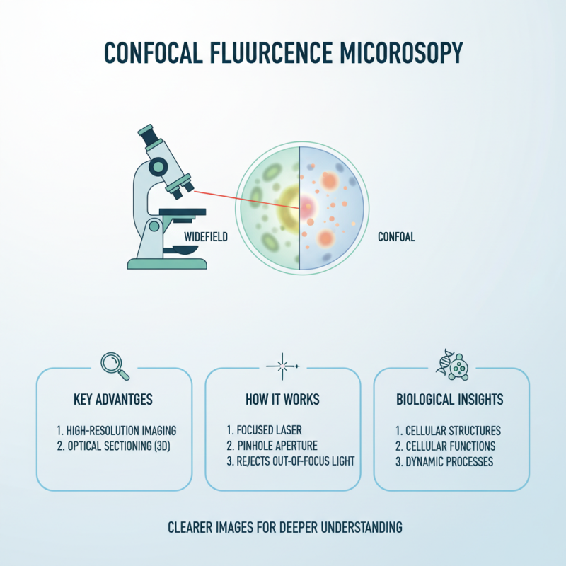
Confocal fluorescence microscopy has transformed biological research in significant ways. This advanced technique allows scientists to visualize structures within cells with remarkable clarity. Equipped with a laser and a pinhole detector, it curates images by focusing on a single plane. The result is stunning resolution, making subtle details more accessible.
Researchers value this method for its ability to reduce background noise. It extracts high-contrast images, even in dense samples. However, this precision comes with challenges. Setting up the equipment can be intricate, often requiring skilled operators. Moreover, sample preparation is crucial, and even minor flaws can result in misleading data.
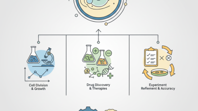
The technique also enables three-dimensional imaging, offering a more profound understanding of cellular interactions. This depth is essential for studying complex processes, such as cell division or signaling pathways. Yet, time constraints can limit comprehensive studies, leaving some questions unanswered. Balancing depth and breadth in research remains a constant challenge for many scientists.
Confocal fluorescence microscopy offers exceptional advantages for biological research, especially in high-resolution imaging. This technique provides detailed insights into cellular structures and functions. By using a focused laser, confocal microscopy eliminates out-of-focus light. This leads to clearer images, enhancing the study of cellular processes.
When capturing images, adjust the laser intensity carefully. Too much light may cause photobleaching, a common issue that can affect your results. Always balance clarity and brightness. Image clarity is crucial for accurate data interpretation. If images appear grainy, review your settings to ensure the optimal resolution.
Another benefit is the ability to perform multi-color imaging. Researchers can label different cellular components with various fluorescent markers. This creates a vivid picture of biological processes. However, overlapping signals may confuse the interpretation. It's essential to choose markers wisely and validate your results.
Experiment with different scanning speeds. Slower speeds can increase resolution but may extend the acquisition time. Time constraints often challenge researchers, so finding the right balance is key. Always consider the trade-offs between quality and efficiency during your experiments.
Confocal fluorescence microscopy stands out in biological research for its enhanced depth perception through optical sectioning. This technique allows scientists to obtain precise images of thick specimens. It achieves this by using a focused laser beam that captures images layer by layer. This creates a three-dimensional effect that is invaluable for understanding complex biological structures.
When examining tissues or cells, the ability to isolate specific planes becomes vital. Researchers can visualize structures buried deep within a specimen without interference from unwanted light. However, despite its advantages, the technique has limitations. Sample preparation can be labor-intensive and requires optimization. Not all fluorophores perform equally well, and some may obscure important details.
Moreover, the data analysis can be challenging. High-resolution images generate substantial amounts of data. This demands significant computational resources and expertise for effective interpretation. While the benefits of enhanced depth perception are clear, the complexities involved mean that researchers must navigate these hurdles to fully utilize this technology.
This chart highlights the top advantages of using confocal fluorescence microscopy in biological research, showcasing aspects such as enhanced resolution, optical sectioning, and improved depth perception, with their corresponding percentage benefits indicated.
Confocal fluorescence microscopy is a powerful tool in biological research. Its ability to improve the signal-to-noise ratio is noteworthy. This enhancement allows researchers to obtain clearer images, which is crucial for analyzing complex biological samples. Higher signal clarity leads to more accurate data collection, making it easier to observe cellular structures and dynamics.
The technique filters out background noise, allowing scientists to focus on the specific signals emitted by fluorescent tags. This specificity is especially important in crowded cellular environments. However, obtaining perfect images remains a challenge. Variations in sample preparation can lead to unexpected noise. Researchers must continually refine their methods to minimize these inconsistencies.
Additionally, while the resolution improvements are significant, artifacts can still appear in images. These artifacts may mislead researchers, prompting incorrect conclusions. Continuous training and practice are essential for fine-tuning this microscopy technique. Embracing these imperfections might actually enhance the learning process, driving further innovations in the field.
Confocal fluorescence microscopy has transformed cellular studies and live-cell imaging. This powerful technique allows researchers to examine cells in real-time. It provides high-resolution images with a significant reduction in background noise. As a result, scientists can focus on specific cellular structures or processes.
In live-cell imaging, this method showcases dynamic cellular activities. For instance, tracking the movement of proteins can reveal cellular pathways. However, achieving optimal imaging conditions can be tricky. Factors like light exposure can affect cell health. Balancing visibility and viability is often a challenge.
Additionally, the use of confocal microscopy opens new doors but has limitations. The complexity of data analysis can overwhelm researchers. Proper training is essential to maximize the technology's potential. Embracing this technique requires both understanding and caution. Observations often lead to more questions than answers, pushing the boundaries of biological research.