10 Best Cell Imaging Systems for Global Buyers?
Choosing among the 10 Best Cell Imaging Systems for Global Buyers requires more than comparing magnification, resolution, or software features. A practical evaluation should connect instrument performance with daily laboratory work. Can the system capture weak fluorescence from living cells without excessive phototoxicity? Does its autofocus remain stable during overnight experiments? Are service engineers available in the buyer’s region? These details often decide whether a powerful platform becomes useful or frustrating.
As cell biologist Jennifer Lippincott-Schwartz has observed, “Seeing is believing, but it is not understanding.” The warning matters. Clear images still require controlled samples, validated protocols, and careful interpretation. This guide examines leading cell imaging platforms through those realities. It considers widefield, confocal, high-content, super-resolution, and live-cell systems. It also reviews workflow speed, environmental control, automation, data handling, and total ownership costs. A system that looks affordable may need expensive objectives, licenses, or maintenance contracts. A premium platform may be excessive for routine endpoint imaging.
Global buyers face additional complications. Voltage standards, import procedures, local training, and replacement-part availability can affect delivery and uptime. These factors are easy to overlook. They should not be.
No ranking can fit every laboratory. Research goals differ. Budgets differ. Even sample preparation changes the result. The comparisons ahead therefore aim to support informed decisions, not promote one universal winner. Some conclusions remain imperfect because specifications and regional support can change. Buyers should confirm current details with manufacturers and independent users before purchasing.
What Is a Cell Imaging System and How Does It Work?
What Is a Cell Imaging System and How Does It Work?
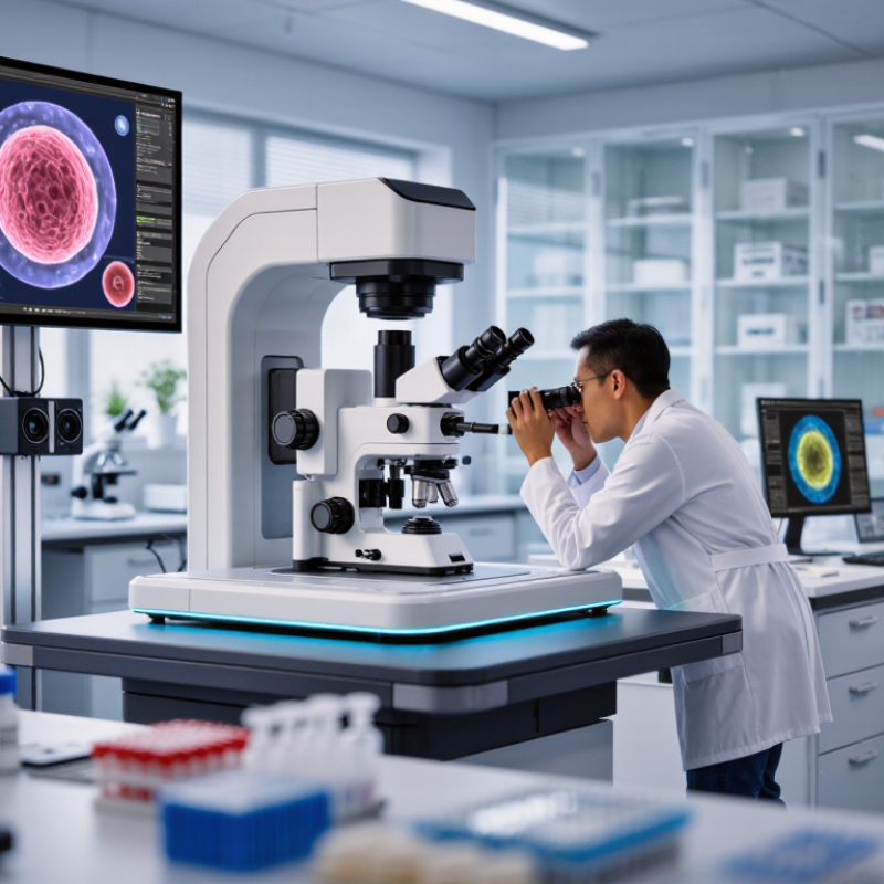
A cell imaging system captures visual information from living or fixed cells. It combines controlled illumination, optical lenses, a camera, and analysis software. Brightfield imaging shows cell shape and density. Fluorescence imaging detects labeled proteins, organelles, or nucleic acids. Some systems also collect images over time, revealing division, migration, and cell death.
The workflow is precise but practical. Cells grow in plates or chambers under controlled temperature and carbon dioxide. Light reaches the sample, and emitted or transmitted signals travel through the objective lens. The camera converts these signals into digital images. Software then measures area, intensity, movement, or object count. Grand View Research reported that the global cell imaging market was valued at approximately USD 2 billion in 2023, with continued growth expected through 2030. This demand reflects wider use in drug discovery, diagnostics research, and cell biology.
Good imaging requires more than expensive hardware. Experienced users standardize exposure time, focus, cell density, and calibration. Excessive light can damage live cells. Poor segmentation can also create confident but false measurements. The 2023 Global R&D Funding Forecast by R&D World highlights continued investment in life-science research, yet funding does not remove experimental uncertainty. A useful system should therefore produce repeatable images, preserve metadata, and support transparent analysis. Automation helps, but it is not perfect. Human review still matters.
Key Features to Compare in Cell Imaging Equipment
When comparing the 10 best cell imaging systems, start with the experiment, not the brochure. A system should match your cell type, culture format, and imaging frequency. Check whether it supports plates, flasks, or specialized chambers. Confirm temperature, humidity, and carbon dioxide control for live-cell work. Small environmental changes can alter cell behavior. They can also damage repeatability.
Optical performance deserves close attention. Compare objective quality, numerical aperture, resolution, field of view, and autofocus stability. A wider field is not always better. It may reduce detail near the edges. Examine brightfield, fluorescence, and transmitted-light options under real sample conditions. Ask how the system handles weak signals, thick samples, and phototoxic exposure. In practical testing, uneven illumination can become a serious problem. It is easy to overlook.
Software can determine daily productivity. Look for automated focusing, stitching, cell counting, confluence measurement, and customizable analysis workflows. Exportable data and clear audit trails support reliable reporting. Confirm that files remain usable without costly restrictions. Also assess calibration routines, maintenance needs, training, and technical support response times. Faster imaging is useful, but only when image quality remains consistent. No system is perfect. A pilot test with your own cells may reveal limitations that specifications hide.
10 Leading Cell Imaging Systems for Global Buyers
Leading cell imaging systems for global buyers must do more than produce sharp pictures. They should capture brightfield, fluorescence, and time-lapse data with consistent focus. In practical laboratory work, a clear workflow matters as much as optical resolution. Operators need simple sample loading, stable illumination, and software that reduces manual adjustments. Small delays become costly when dozens of plates wait nearby. Keep it practical.
Experienced users also examine calibration tools, image quality controls, and data traceability. Reliable systems should preserve acquisition settings, record user actions, and export files in widely accepted formats. These features support repeatable experiments across research centers, hospitals, and contract laboratories. A responsive service network matters too, especially when replacement parts cross borders. Buyers should check installation training, warranty terms, local language support, and electrical compatibility before signing an order.
Global purchasing brings less obvious challenges. A compact system may suit a small laboratory, while a high-throughput platform may need more space, cooling, and technical staffing. Some platforms advertise advanced automation, yet daily users may find the software difficult to learn. That risk deserves honest testing. Ask for representative samples, not only polished demonstrations. I would compare live-cell stability, autofocus performance, cleaning requirements, and five-day operating costs. The highest resolution is not always the most useful result. A system that researchers can operate correctly every day may deliver stronger evidence than a more powerful instrument left underused.
Applications Across Research, Healthcare, and Drug Development
10 Best Cell Imaging Systems for Global Buyers?
Cell imaging now supports research, healthcare, and drug development. In research laboratories, high-content platforms track cell shape, movement, protein location, and viability. These details can reveal biological changes before they become visible in ordinary microscopy. The 2024 IQVIA Institute report recorded more than 20,000 active clinical development programs worldwide. That pipeline increases demand for faster, reproducible imaging data. Yet, more images do not always mean better science.
Healthcare applications are becoming more practical. Imaging systems assist digital pathology, blood-cell analysis, tissue assessment, and disease monitoring. The World Health Organization’s World Health Statistics 2024 reports that noncommunicable diseases caused about 74% of global deaths in 2021. This burden makes earlier, measurable cellular signals valuable. Drug developers also use live-cell imaging for phenotypic screening, toxicity studies, and dose-response analysis. Small changes matter. However, uneven sample quality, fluorescent bleaching, and algorithm bias can weaken results. Buyers should examine validation records, image-export options, service coverage, and data security across regions.
Tips: Compare systems using the same sample set. Request raw images, not only processed figures. Check illumination stability over long experiments. Ask whether analysis tools can be independently audited. A cheaper instrument may create higher costs through repeated tests, but expensive hardware is not automatically more reliable. Teams should document failed runs, too. That uncomfortable record often exposes workflow problems before they affect clinical or development decisions.
How to Select the Right System for Your Laboratory Needs
Selecting a cell imaging system starts with your laboratory’s daily questions, not a ranked list of popular instruments. Define the samples, dyes, magnification, throughput, and resolution you truly need. A system for fixed-cell morphology may not suit live-cell observation. I have seen laboratories overpay for advanced features used only twice each year.
Check the workflow in practical detail. Can a technician load a plate without disturbing samples? Does the software measure confluence, fluorescence intensity, or cell movement consistently? Ask for raw image access, export formats, and audit trails. These details matter when results support regulated research or shared projects. Test representative samples, including dim signals and crowded fields. Clean images can hide weak performance.
Budget beyond the purchase price. Include installation, service response, training, replacement parts, and compatible consumables. Confirm electrical requirements, data storage, cybersecurity controls, and local compliance obligations before ordering internationally. A compact system may fit beside the incubator, while a larger platform could require room redesign. That is easy to miss.
Request an application-based demonstration with your own protocol. Compare signal-to-noise, autofocus reliability, acquisition time, and analysis repeatability. Let several users evaluate the interface. Their feedback may disagree. That is useful. Document every limitation, because the most honest selection often comes from knowing what the system cannot do.