The Best 10 Techniques in Cell Imaging You Should Know?
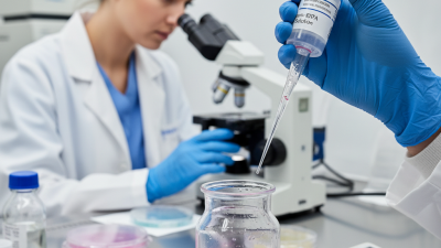
Cell imaging is a vital technique in biomedical research. It allows scientists to visualize cells in real-time. Powerful imaging techniques shed light on cellular processes and structures. However, mastering these techniques can be challenging. The intricacies of cell imaging methods can lead to confusion and misinterpretation.
The best techniques can enhance your research. Each method has its strengths and weaknesses. For example, fluorescence microscopy is popular but can suffer from photobleaching. Electron microscopy provides stunning detail but may require complex sample preparation. Understanding these trade-offs is essential for effective cell imaging.
As we explore the best techniques, we must acknowledge their limitations. No single method is perfect. Factors like cost, complexity, and resolution all play a role. Embracing these imperfections can lead to innovative solutions and improved research outcomes. In this ever-evolving field, keeping abreast of new developments is crucial. These ten techniques stand at the forefront, shaping the future of cell imaging.
Overview of Cell Imaging Techniques
Cell imaging techniques are essential for understanding biological processes. They allow researchers to visualize cells in real-time. This overview highlights several prominent methods used in laboratories today.
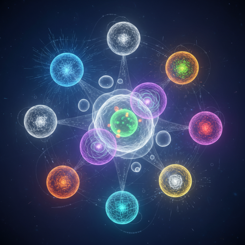
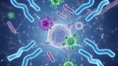
Fluorescence microscopy is popular for studying cell structures. It uses fluorescent dyes to label specific proteins. This method provides high contrast images. However, it can sometimes lead to photobleaching. Researchers must be cautious with dye selection. Live-cell imaging techniques offer another layer of understanding. They help observe dynamic processes within cells. Yet, keeping cells alive during prolonged observation is a challenge.
Another technique is electron microscopy. This method provides very high-resolution images. It reveals fine details of cell ultrastructure. Nonetheless, sample preparation can alter cell morphology. Researchers often need to reflect on how artifacts impact results. When combining multiple imaging techniques, it’s vital to analyze the data accurately. Sources of error must be considered to avoid misinterpretations.
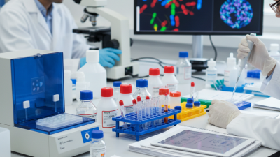
Tips: Always calibrate equipment before use. Take control images for comparison. Stay organized with data management to maintain reproducibility.
The Best 10 Techniques in Cell Imaging You Should Know
| Technique | Description | Advantages | Limitations |
|---|---|---|---|
| Fluorescence Microscopy | Uses fluorescence to visualize structures within cells. | High sensitivity and specificity, can be used in live cell imaging. | Photobleaching and background noise can affect results. |
| Confocal Microscopy | Offers improved resolution by using a pinhole to eliminate out-of-focus light. | Provides 3D images and can capture dynamic processes. | More expensive and can be complex to operate. |
| Electron Microscopy | Uses electron beams to obtain high-resolution images of cell ultrastructure. | Exceptional resolution, providing detailed images at the nanometer scale. | Sample preparation is complex and cells must be dead. |
| Phase Contrast Microscopy | Enhances the contrast of transparent specimens to view live cells. | Can observe live cells without staining. | Lower resolution compared to fluorescence and confocal microscopy. |
| Live Cell Imaging | Real-time observation of living cells using various imaging techniques. | Allows for dynamic studies of cellular processes. | Limited time frame and requires specialized equipment. |
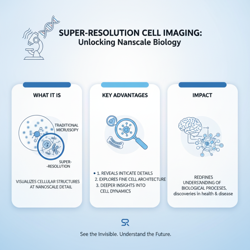
| Super-Resolution Microscopy | Methods such as STORM and PALM break the diffraction limit of light microscopy. | High spatial resolution, revealing details down to nanometers. | Complex techniques and may require extensive computational power. |
| Total Internal Reflection Fluorescence (TIRF) Microscopy | Illuminates a small region of the sample, ideal for studying membrane dynamics. | Excellent for low-background imaging of live cells. | Limited to the interface of the sample and substrate. |
| Atomic Force Microscopy (AFM) | Uses a cantilever with a sharp tip to map the surface of a sample on the nanoscale. | High-resolution topographic data. | Slow imaging speed and requires specialized conditions. |
| Multiphoton Microscopy | Uses two or more photons to excite fluorescent markers at greater depths within tissues. | Deeper tissue imaging with reduced phototoxicity. | Higher equipment costs and more complex setup. |
| Flow Cytometry | Analyzes the physical and chemical characteristics of cells in a fluid suspension. | High-throughput analysis of multiple parameters. | Requires dissociation of cells from tissue. |
Related Posts
-
2026 Best Secondary Antibodies for Your Research Needs?
-
What is a cell proliferation assay and how is it used?
-
How to Use Trypsin EDTA for Cell Culture and Tissues?
-
Top 10 Facts About IGG Antibody That Everyone Should Know?
-
What is 3D Cell Culture and How Does It Work?
-

Why Are Monoclonal Antibodies Important for Disease Treatment?