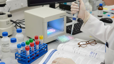
Choosing the top 10 minimal residual disease tests requires more than listing familiar technologies. It requires context. A blood tube, a bone marrow aspirate, or a tissue sample can produce very different answers. Disease type, treatment stage, specimen quality, and laboratory expertise all influence the result.
As hematopathologist Michael J. Borowitz has stated, “Minimal residual disease is the most important prognostic factor in acute lymphoblastic leukemia.” His observation explains why clinicians monitor disease signals after treatment, even when standard microscopy shows remission. A patient may appear clinically well while a highly sensitive test detects a small remaining population of malignant cells.
This guide examines ten widely used approaches, including multiparameter flow cytometry, quantitative PCR, digital PCR, next-generation sequencing, and circulating tumor DNA analysis. Each method has strengths and limitations. Some offer rapid results. Others provide deeper sensitivity or better molecular specificity. None is flawless. A negative result does not always mean that every malignant cell has disappeared.
The ranking is therefore practical, not absolute. A test may be excellent for measurable disease in leukemia but less useful for a solid tumor. Sample handling matters. So does the reporting threshold. Even experienced teams can interpret borderline findings differently.
Readers should use this overview to understand test selection, analytical sensitivity, clinical relevance, and common sources of uncertainty. Treatment decisions must remain with qualified healthcare professionals who can interpret results alongside pathology, imaging, symptoms, and the patient’s full medical history.

Minimal residual disease (MRD) means cancer cells remain after treatment, even when scans show no visible tumor. These cells may be too few for routine imaging or standard blood tests. MRD testing can detect traces in blood, bone marrow, or tissue. It may help doctors estimate relapse risk and adjust follow-up care.
The “top 10” MRD tests are not a universal ranking. Available methods include flow cytometry, polymerase chain reaction, digital PCR, next-generation sequencing, circulating tumor DNA analysis, and specialized tissue tests. Their accuracy depends on cancer type, sample quality, treatment history, and the genetic features of each patient’s disease. MRD is useful. It is not a crystal ball. A negative result does not always mean every cancer cell has disappeared.
Tips: Ask what sample the test needs and why it suits your cancer. Request the detection limit and the meaning of a negative result. Confirm whether results should be compared with earlier samples. Timing matters. A test taken too soon may not reflect treatment response clearly. Discuss unexpected findings with a qualified oncology team, because laboratory results require clinical context. Even experienced clinicians may revise their interpretation when new evidence appears.
Minimal residual disease (MRD) refers to a very small number of cancer cells that may remain after treatment and cannot usually be detected by routine imaging or standard laboratory tests. The chart compares commonly used or actively studied MRD testing approaches by their approximate analytical detection level.
Values show representative orders of magnitude reported for assay sensitivity, expressed as the detectable fraction of abnormal cells or molecules. Actual performance varies by cancer type, sample quality, target biology, laboratory protocol, and disease burden. A lower value indicates greater analytical sensitivity; this is not a clinical ranking.
Minimal residual disease tests work by searching for cancer cells after treatment, when routine microscopy may look normal. The leading ten approaches include multiparameter flow cytometry, allele-specific qPCR, digital PCR, next-generation sequencing, RNA fusion testing, FISH, conventional cytogenetics, mass spectrometry, PET/CT, and MRI. Each detects a different signal. Flow cytometry identifies abnormal surface proteins on individual cells. PCR amplifies a known genetic sequence. Digital PCR divides samples into thousands of reactions, improving rare-target counting. Sequencing reads mutations or rearranged immune genes across many DNA fragments.
Sensitivity varies sharply. European LeukemiaNet guidance reports that flow cytometry can often detect one abnormal cell among 10,000, while molecular methods may reach one among 100,000 or more. The International Myeloma Working Group recognizes imaging as complementary, not interchangeable, because PET/CT shows active lesions rather than every malignant cell. Timing matters too. A marrow sample taken too early can reflect treatment injury. Sampling site matters.
Blood is easier to collect.
Bone marrow can be more informative.
However, no test is universally best. Clonal evolution may remove the original target, creating a false-negative result. Hemodilution can also weaken marrow findings. A 2023 professional laboratory review emphasized analytical sensitivity, specimen quality, and standardized reporting as major reliability factors. In practice, clinicians compare test results with disease type, treatment response, and patient symptoms. The numbers help, but they do not replace judgment.
Residual cancer cells can hide after treatment, even when scans look clear. The ten leading detection methods are bone-marrow morphology, multiparameter flow cytometry, allele-specific PCR, digital PCR, targeted next-generation sequencing, whole-genome sequencing, circulating tumor DNA analysis, circulating tumor-cell counting, methylation testing, and functional imaging with PET or MRI. They answer different questions.
Sensitivity varies sharply. International Myeloma Working Group guidance reports that next-generation sequencing and advanced flow cytometry may detect roughly one abnormal cell among 100,000 or one million normal cells. PCR can reach similar levels when a patient’s molecular marker is known. EuroFlow technical reports also show that sample quality strongly affects flow-based results. A delayed marrow sample may weaken the signal.
Blood-based tests are less invasive. ctDNA can sometimes reveal molecular relapse before radiographic progression, often by several months in research cohorts. However, low tumor shedding can produce a false negative. PET and MRI show active or anatomical disease, not always microscopic cells. Methylation assays remain promising but need broader clinical validation. No single method fits every cancer.
Test interpretation needs clinical experience. Results depend on disease type, specimen timing, treatment response, and laboratory quality controls. A negative result is reassuring, not absolute. Some reports also compare different thresholds, which makes cross-study claims imperfect and worth questioning.
What Are the Top 10 Minimal Residual Disease Tests?
The leading MRD approaches include multiparameter flow cytometry, allele-specific qPCR, digital PCR, conventional NGS, error-corrected NGS, RNA sequencing, fusion-transcript PCR, methylation assays, circulating tumor DNA sequencing, and imaging-supported assessment. Their performance differs sharply. Flow cytometry usually detects one abnormal cell among 10,000 to 100,000 cells. It is practical for leukemia and myeloma, but depends on viable cells and expert interpretation. Allele-specific qPCR can reach about 10^-5 sensitivity when a suitable molecular marker exists. Digital PCR may improve precision at very low disease levels, especially during treatment monitoring.
NGS can identify several mutations at once and may reach 10^-5 or deeper with error correction. It supports leukemia, lymphoma, myeloma, and selected solid tumors. However, clonal hematopoiesis can create misleading signals. RNA-based assays are useful when disease-specific transcripts are stable. Methylation testing may help classify tumor-derived DNA, although clinical validation remains uneven. Circulating tumor DNA is attractive for solid tumors because blood collection is less invasive. Its sensitivity falls when tumors shed little DNA. Imaging can reveal disease outside the sampled compartment, but it cannot replace molecular testing.
Specificity matters as much as sensitivity. A test may detect tiny signals yet struggle to prove they represent active cancer. Sample timing, tissue location, tumor biology, and laboratory quality all influence results. A negative result is not always reassuring. Sometimes, it reflects an inadequate sample. The most reliable clinical decision combines validated testing, repeat measurements, and patient-specific context. No single method wins every case.
Comparative overview of commonly used and emerging MRD technologies in hematologic and solid-tumor settings.
| Rank | MRD Test | Primary Detection Principle | Typical Analytical Sensitivity | Typical Specificity | Best-Suited Clinical Applications | Main Strengths | Important Limitations |
|---|---|---|---|---|---|---|---|
| 1 | Next-Generation Sequencing of Immunoglobulin or T-Cell Receptor Rearrangements | Tracks patient-specific clonal immune-receptor sequences in malignant B or T cells. | 10−5 to 10−6 | >99% when the baseline clone is correctly identified | Acute lymphoblastic leukemia, multiple myeloma, chronic lymphocytic leukemia, and other B- or T-cell neoplasms. | Very high sensitivity; provides a quantitative result; useful for serial monitoring. | Requires an adequate diagnostic specimen and an identifiable baseline clonotype; clonal evolution or sampling variation may affect interpretation. |
| 2 | Multiparameter Flow Cytometry | Identifies abnormal antigen-expression patterns on individual cells using multiparameter immunophenotyping. | 10−4 to 10−5 | Approximately 95% to >99%, assay-dependent | Acute myeloid leukemia, acute lymphoblastic leukemia, multiple myeloma, and selected lymphomas. | Rapid turnaround; broadly applicable; does not always require a patient-specific molecular target. | Dependent on specimen quality, hemodilution, antibody panels, operator expertise, and the stability of the abnormal phenotype. |
| 3 | Allele-Specific Quantitative PCR | Amplifies a patient-specific fusion transcript, rearrangement, or mutation using designed primers and probes. | 10−4 to 10−5 | Approximately 95% to >99% | BCR::ABL1-positive leukemia, selected acute leukemias, and diseases with a stable, trackable molecular lesion. | Well-established workflows; relatively fast; quantitative and highly sensitive for validated targets. | Requires a suitable molecular marker and patient-specific assay design; not applicable to every case. |
| 4 | Digital PCR or Droplet Digital PCR | Partitions DNA or RNA molecules into thousands of reactions and uses endpoint fluorescence to quantify rare targets. | 10−4 to 10−5 | >99% for a well-validated target | Monitoring recurrent mutations, fusion transcripts, and other known molecular markers in leukemia and lymphoma. | High precision at low target concentrations; less dependent on standard curves; useful for longitudinal testing. | Usually limited to known targets; primer or probe design and background DNA quality strongly influence performance. |
| 5 | Error-Corrected Targeted NGS | Uses molecular barcodes, duplicate consensus reads, and bioinformatic error suppression to detect low-frequency variants. | 10−4 to 10−6 | >99% after validated error suppression | Mutation-defined acute myeloid leukemia, myelodysplastic syndromes, myeloma, lymphoma, and other molecularly trackable cancers. | Can evaluate multiple genes or mutations in one assay; supports broader molecular profiling. | Requires rigorous validation; clonal hematopoiesis can complicate interpretation; mutation clearance may not always equal eradication of malignant cells. |
| 6 | Reverse-Transcription Quantitative PCR for Fusion Transcripts | Measures disease-associated RNA transcripts generated by recurrent chromosomal rearrangements. | 10−4 to 10−5 | Approximately 95% to >99% | Leukemias with stable recurrent fusions, including kinase and transcription-factor rearrangements. | Very sensitive for the appropriate fusion; suitable for standardized serial monitoring. | RNA is less stable than DNA; transcript levels may vary by disease state; only applicable when the relevant fusion is present. |
| 7 | Molecular MRD by Conventional Targeted NGS | Detects residual disease-associated sequence variants using high-depth sequencing without advanced error-correction methods. | 10−3 to 10−4 | Approximately 95% to 99%, depending on depth and filtering | Broad screening and mutation tracking in leukemia, lymphoma, myeloma, and selected solid tumors. | Can assess several potential markers simultaneously; useful when a single dominant marker is not available. | Low-frequency sequencing errors can produce false positives; residual mutations may represent nonmalignant clonal hematopoiesis. |
| 8 | Fluorescence In Situ Hybridization | Uses fluorescent probes to identify disease-associated chromosomal rearrangements, amplifications, or deletions in individual cells. | 10−2 to 10−3 | Approximately 95% to >99% for a clearly defined abnormality | Plasma-cell neoplasms, lymphomas, leukemias, and tumors with characteristic cytogenetic abnormalities. | Works on non-dividing cells; can confirm specific structural or copy-number abnormalities. | Generally less sensitive than flow cytometry or molecular assays; requires a known target and adequate cell counting. |
| 9 | Plasma Circulating Tumor DNA NGS | Analyzes tumor-derived DNA fragments circulating in plasma using targeted or broader sequencing approaches. | Approximately 10−4 to 10−6 in optimized assays | Approximately 95% to >99%, assay- and tumor-dependent | Emerging MRD monitoring after treatment of colorectal, breast, lung, and other solid tumors. | Minimally invasive; may reflect disease throughout multiple anatomical sites; useful when tissue sampling is difficult. | Not yet equally standardized across cancers; low tumor shedding, clonal hematopoiesis, and limited plasma DNA can reduce accuracy. |
| 10 | Plasma Circulating Tumor DNA Digital PCR | Quantifies a predefined tumor mutation or methylation marker in cell-free DNA using highly partitioned PCR reactions. | 10−3 to 10−5 | Approximately 95% to >99% for a validated target | Focused postoperative or post-treatment surveillance of solid tumors with a known molecular alteration. | Highly quantitative; relatively rapid; requires less sequencing infrastructure than NGS. | Limited to known alterations; plasma shedding varies substantially by tumor type, size, site, and treatment status. |
Doctors do not choose the “best” minimal residual disease test in isolation. They match the test to the cancer, sample, treatment stage, and clinical question. Common options include multiparameter flow cytometry, quantitative PCR, digital PCR, next-generation sequencing, RNA sequencing, error-corrected sequencing, circulating tumor DNA analysis, bone marrow morphology, PET-CT, and MRI. Each measures a different signal.
In multiple myeloma, the 2022 International Myeloma Working Group consensus supports marrow-based sequencing or flow testing, while imaging can reveal disease outside the marrow. A negative marrow result may therefore miss a distant lesion.
Interpretation requires context. Doctors review treatment timing, specimen volume, laboratory quality controls, and the patient’s earlier tumor profile. They also compare trends, not one isolated number.
A low-level signal can reflect true disease, technical noise, or clonal change. The College of American Pathologists emphasizes validated thresholds and documented quality assurance for reliable MRD reporting. Still, these tests are not perfect.
I would question any result that conflicts sharply with symptoms, imaging, or blood counts. A careful discussion is often more useful than a dramatic percentage.