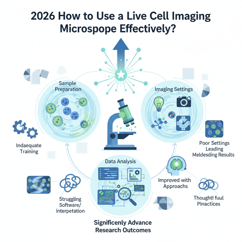
Live cell imaging microscopy has transformed biological research. Dr. Alice Green, a renowned expert in cell biology, once stated, "This technology reveals cellular processes in real time." Understanding how to use a live cell imaging microscope effectively is crucial for researchers.
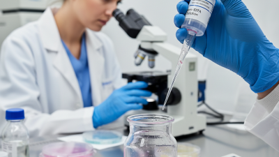
The key factors include sample preparation, imaging settings, and data analysis. Proper sample preparation ensures that cells remain healthy during observation. Adjusting imaging settings enhances clarity and captures dynamic events. Yet, researchers often face challenges. Inadequate training or poor settings can lead to misleading results.
Additionally, analyzing the obtained data is complex. Many researchers may struggle with the software or interpretation of images. Reflection on these challenges can lead to improved practices. A live cell imaging microscope offers remarkable insights, but it requires careful handling and thoughtful approaches. Embracing these aspects can significantly advance research outcomes.
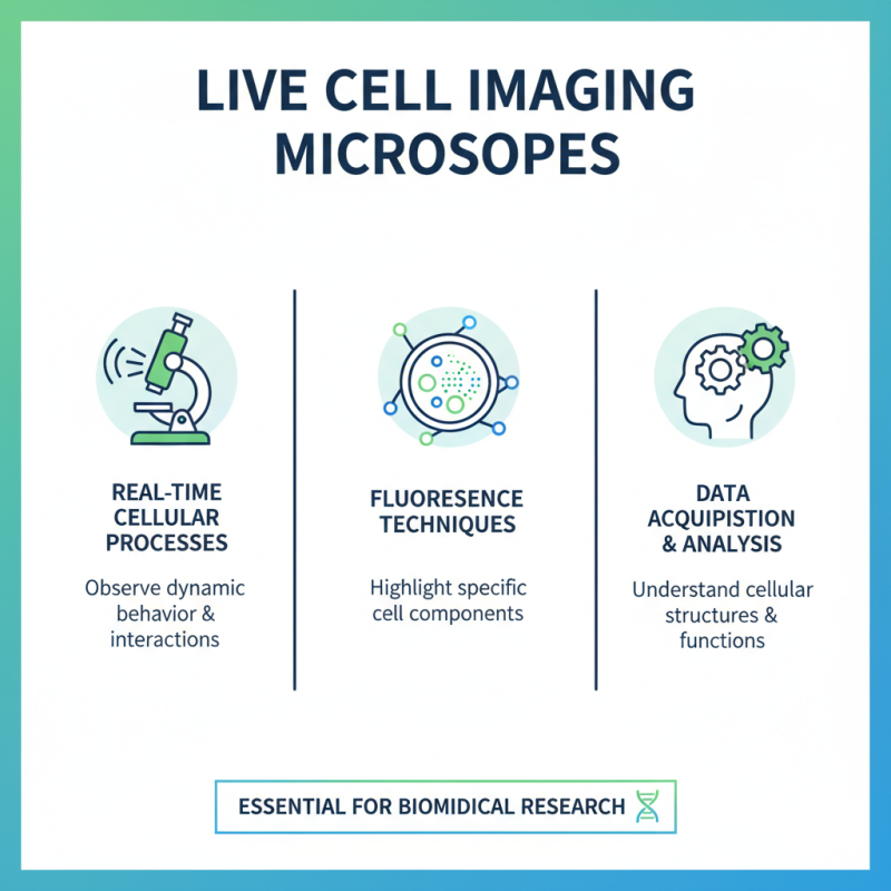
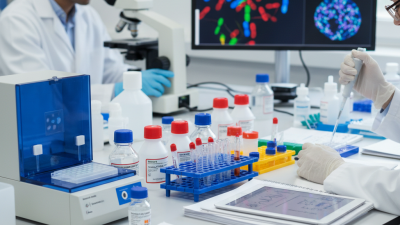
Live cell imaging microscopes are powerful tools for studying cellular processes in real time. These microscopes allow researchers to visualize live cells, observing their behavior under various conditions. By using specific techniques such as fluorescence, scientists can highlight certain cell components. This illumination reveals dynamic interactions and cellular structures. Understanding how to operate this equipment is essential for obtaining meaningful data.
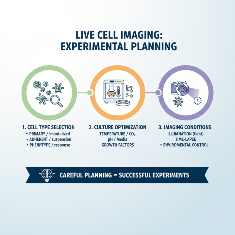
However, challenges often arise during imaging. Maintaining cell viability is crucial. Even slight disturbances can lead to artifacts in the data. Temperature fluctuations can also affect results. Therefore, equipment calibration and environmental control must be considered. Researchers might find themselves grappling with these aspects while trying to optimize their images. This means regular adjustments and vigilance are necessary.
Moreover, interpreting the data is not straightforward. Misinterpretations can stem from noise or overlapping signals. It’s easy to get lost in complex imaging results. Careful analysis is required to validate findings. Thus, researchers should continuously reflect on their methodologies. Striving for improvement in imaging techniques can lead to better experimental outcomes in the long run.
When selecting a live cell imaging microscope, certain key features are essential for effective use. High-resolution imaging capability is crucial. This allows researchers to observe cellular dynamics in great detail. A compact and stable design is also essential, which minimizes movement and vibrations during imaging. Live cell systems must maintain environmental conditions like temperature and CO2 levels. This ensures cells remain viable during observations.
Another important aspect is the availability of multi-channel imaging. This feature enables the simultaneous observation of different signals within the same sample. Proper software integration is vital for easy operation. User-friendly interfaces simplify the complexity of analysis and data handling. However, while these features are critical, the learning curve for new users may be steep. It's important to allow for practice and adjustment.
Finally, consider the microscope’s versatility. Can it adapt to various imaging techniques? Flexibility may enhance your research options. Yet, be cautious. A microscope that attempts to do too much may compromise specific capabilities. Striking the right balance is key. Remember, each laboratory is different. Reflect on your unique needs to make the best choice.
Setting up a live cell imaging microscope requires careful attention to detail. Begin by ensuring that the microscope is clean and calibrated. A small error in calibration can lead to significant deviations in data. According to a recent industry report, improper setup can reduce imaging accuracy by over 30%. This emphasizes the need for a meticulous approach.
Lighting is crucial in live cell imaging. Adjust the light source to minimize phototoxicity while ensuring adequate illumination. Overexposure can harm delicate cells, especially in long-term studies. Aim for a balanced intensity, which helps maintain cell viability for hours, sometimes days. In fact, studies reveal that optimal light conditions improve imaging quality by about 25%.
Finally, familiarizing yourself with software settings is essential. Many users overlook this aspect. A tangled setup can lead to missed observations. Regularly updating software ensures compatibility and efficiency. It's easy to assume everything is fine, but periodic checks can reveal areas for improvement. These adjustments can enhance both the reliability of your findings and the longevity of your samples.
| Step | Description | Time Required | Materials Needed |
|---|---|---|---|
| 1 | Select the appropriate microscope setup based on your samples and experiment. | 20 minutes | Microscope, digital camera |
| 2 | Prepare live samples according to the protocol specific to the cells. | 30 minutes | Culture media, cell lines |
| 3 | Calibrate the microscope settings including focus, exposure time, and illumination. | 15 minutes | Calibration slides |
| 4 | Load the live samples into the imaging chamber, ensuring minimal disturbance. | 10 minutes | Imaging chamber, lid |
| 5 | Take images at defined time intervals and conditions to monitor live cell behavior. | Variable | Computer with imaging software |
| 6 | Analyze the images using appropriate software to quantify cell responses. | 40 minutes | Analysis software |
Live cell imaging is a powerful technique used in biological research. To conduct effective experiments, careful planning is essential. Start by selecting the right cell type for your study. Consider the specific characteristics you wish to observe. Different cells respond differently to imaging conditions. Ensure you optimize your culture conditions. Pay attention to factors like temperature, pH, and nutrient availability.
Another vital aspect is the imaging environment. Minimize light exposure to reduce photobleaching. Adjust your microscope settings, including brightness and contrast, for the best image quality. Be prepared for adjustments during your experiment. Sometimes, settings need real-time modification based on the observed results. Don't be disheartened by unexpected outcomes; they can lead to new questions.
Documentation is crucial throughout the process. Record your methods and observations meticulously. This information is invaluable for future experiments. Reflect on your techniques and outcomes regularly. Ask yourself, could the cells have reacted differently? Evaluate your experimental design and make changes where needed. Continuous improvement is key to mastering live cell imaging.
Live cell imaging provides fascinating insights into cellular processes. However, analyzing the resulting data can be quite challenging. One crucial technique is tracking cellular movements over time. Using software can help visualize these changes. It allows for precise measurement of cell velocity and migratory patterns. But, this requires careful calibration. Any errors can lead to misleading results.
Another vital aspect of data analysis is fluorescence intensity quantification. This technique helps in understanding protein expression levels. However, fluctuations in signal can occur. Background noise might distort data interpretation. Analysts must be vigilant in differentiating between actual signals and noise. Implementing controls is essential, but even then, doubt can linger.
Data visualization is key in presenting findings. Graphs and heat maps can simplify complex data sets. Yet, sometimes these visual tools can be misleading. They may fail to capture the full picture. It is important to reflect on the limitations of each analysis approach. A thorough understanding of each technique enhances the accuracy of results. Balancing rigor with flexibility in methodology is crucial for meaningful insights.