2026 How to Use Goat Anti Rabbit HRP for Effective Immunohistochemistry?

Immunohistochemistry (IHC) has evolved as a crucial technique in biological research. The use of goat anti rabbit HRP is prominent among various methods. Dr. Emily Thompson, a leading expert in IHC, emphasizes, "The choice of secondary antibodies greatly influences the clarity of imaging." This highlights the importance of selecting the right tools for successful applications.

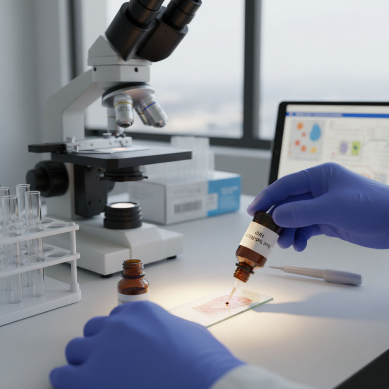
Goat anti rabbit HRP offers excellent specificity and sensitivity. When applied correctly, it significantly improves staining outcomes. It can enhance the visualization of rabbit primary antibodies in tissue samples. This leads to better insights into pathological conditions. However, not all applications yield the same results. The quality of antibodies and protocols must be carefully assessed.

Many researchers face challenges while using goat anti rabbit HRP. Sometimes, background staining occurs, which obscures results. Proper controls are vital to mitigating these issues. Uncertainties about dilution factors and incubation times can also impact outcomes. Thus, ongoing experimentation and critical evaluation are necessary to refine techniques.

2026 How to Use Goat Anti Rabbit HRP for Effective Immunohistochemistry?

Understanding Goat Anti-Rabbit HRP and Its Role in Immunohistochemistry

2026 How to Use Goat Anti Rabbit HRP for Effective Immunohistochemistry?

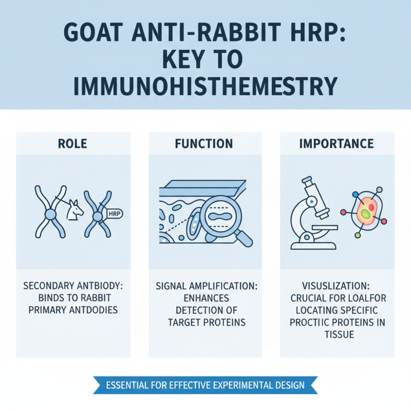
Goat anti-rabbit HRP is a vital reagent in immunohistochemistry. It works as a secondary antibody, enhancing the detection of primary antibodies in tissue samples. This tool is essential for visualizing the presence of specific proteins. By binding to rabbit antibodies, it amplifies the signal, enabling clearer results. Understanding its role helps in designing effective experiments.

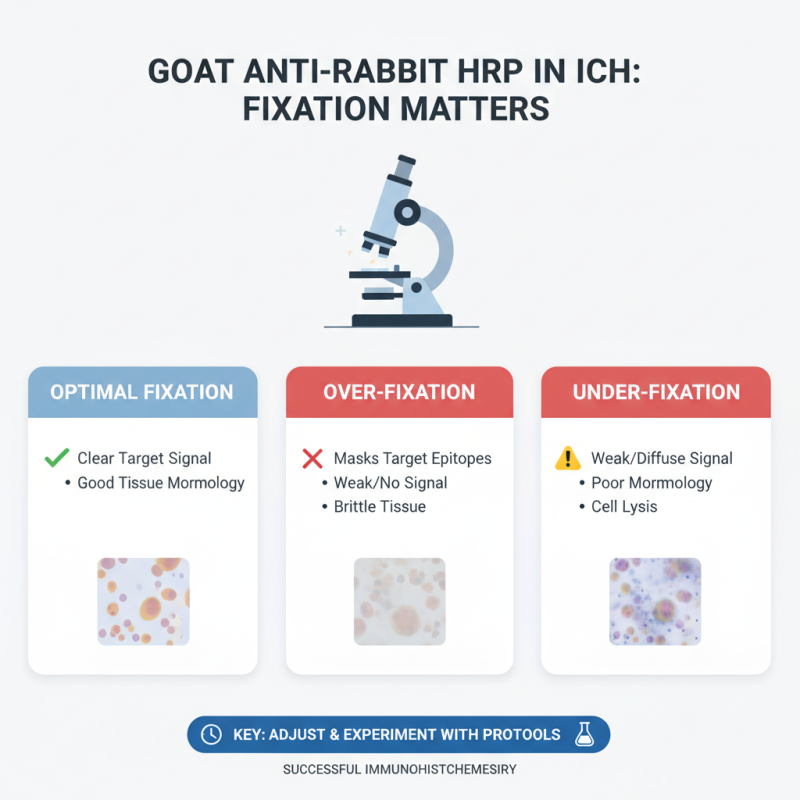
When using goat anti-rabbit HRP, proper dilution is key. A common practice is to test different dilutions. This might seem tedious, but it helps find the optimal concentration. Too much reagent can lead to high background noise. A good starting point is a 1:500 dilution and adjusting based on the results.

Tips: Always include controls in your experiments. This can guide you in interpreting your results accurately. Make sure your tissue samples are well-prepared to minimize variability. It’s crucial to take notes during the process. Reflecting on what worked and what didn’t can improve future experiments. Keep in mind, outcomes can vary based on numerous factors like tissue type and handling.

Choosing the Right Protocol for Immunohistochemical Staining

Choosing the right protocol for immunohistochemical staining is crucial for obtaining reliable results. Proper selection can enhance specificity and signal intensity. Studies show that goat anti-rabbit HRP conjugates provide excellent sensitivity. They increase detection limits, enabling visualization of low-abundance proteins. This is especially valuable in clinical research.

The protocol should match the tissue type and target antigens. It’s essential to optimize conditions like antibody dilution and incubation time. According to recent data, inappropriate dilutions can lead to background noise. Troubleshooting is an integral part of this process. A poorly tuned protocol can yield inconsistent results. In some cases, signal loss might occur due to overblocking.

Furthermore, using proper controls can guide protocol adjustments. Negative controls help identify non-specific binding. Data indicate that using primary antibodies in a concentration range of 1:100 to 1:1000 is effective. However, this may vary among experiments. Monitoring these variables can make a significant difference. Every detail matters when striving for accuracy in immunohistochemistry.

2026 How to Use Goat Anti Rabbit HRP for Effective Immunohistochemistry? - Choosing the Right Protocol for Immunohistochemical Staining

Protocol Step Description Recommended Time Notes
Tissue Preparation Fixation and embedding of tissue samples. 1-2 hours Use formalin for fixation.
Sectioning Cutting tissue into thin sections for microscopic examination. 30 minutes Maintain a thickness of 4-5 micrometers.
Deparaffinization Removal of paraffin wax from tissue sections. 15 minutes Use xylene or a suitable clearing agent.
Rehydration Gradual rehydration of tissue sections in alcohol. 10 minutes Use 100%, 95%, and 70% ethanol.
Blocking Blocking non-specific binding sites. 1 hour Use serum from the same species as the secondary antibody.
Primary Antibody Incubation Incubation with Goat Anti-Rabbit HRP antibody. 1-2 hours Dilution factors vary based on antibody.
Washing Removal of unbound antibodies. 5-10 minutes Use phosphate-buffered saline (PBS).
Secondary Antibody Incubation Incubation with secondary antibody conjugated to HRP. 1 hour Ensure compatibility with primary antibody.
Detection Application of HRP substrate for visualization. 5-15 minutes Monitor color development microscopically.

Preparing Tissue Samples for Effective Antibody Binding

Preparing tissue samples is crucial for effective antibody binding in immunohistochemistry. Start by meticulously fixing the tissue. Use a formaldehyde solution for proper preservation. This step minimizes degradation and retains cellular structures. Ensure that the fixation time is optimal; over-fixing can obscure antigen sites. Aim for clarity, but avoid extremes.

Next, consider the embedding process. Choose an appropriate medium. This step supports the tissue during section cutting. If the embedding is inconsistent, it can impact the quality of your sections. Cut thin slices—ideally 4-6 micrometers thick. Thicker sections may trap air bubbles, which hinder antibody penetration. Balance is key here; too thin, and you risk losing important structural details.

Finally, pay attention to deparaffinization for fixed tissues. Use xylene followed by graded alcohols to ensure complete removal of the embedding medium. Any remnants can impede antibody access. Rinse thoroughly in phosphate-buffered saline to prepare for the next steps. Adjustments may be necessary during this process, as everyone's technique may differ slightly. It's essential to stay vigilant and reflect on your methods to improve results.

Optimizing Goat Anti-Rabbit HRP Application Techniques

2026 How to Use Goat Anti Rabbit HRP for Effective Immunohistochemistry?

When working with Goat Anti-Rabbit HRP, attention to detail is crucial for successful immunohistochemistry. The choice of tissue fixation greatly influences staining effectiveness. Over-fixation can mask targets. Under-fixation may lead to weak signal. Adjusting fixation time is key. Experimenting with different fixation protocols can yield varying results.

Another important factor is the dilution of the antibody. Each tissue type may respond differently. A common starting point is to dilute the antibody 1:200. However, some tissues may require higher or lower concentrations. Testing different dilution factors on a control tissue is advisable. This trial and error can optimize staining intensity.

Blocking is equally significant. Adequate blocking prevents non-specific binding. It usually requires 1-2 hours at room temperature. Yet, longer blocking may be beneficial in some cases. Pay attention to background staining, as this can detract from specific signals. Finding the right balance may take time and patience.

Analyzing Results: Interpreting Staining Outcomes in Tissue Sections

Interpreting staining outcomes in immunohistochemistry is crucial for accurate analysis. The staining pattern can reveal important information about protein expression in tissue sections. This information is essential for researchers studying diseases and biological processes.

Analyzing results requires attention to detail. Staining intensity can vary significantly. Some areas may appear dark, while others are lighter. It’s important to assess whether this reflects true biological variability or if it’s due to technical issues. Observing controls can provide context for interpreting experimental results.

Artifacts can complicate results. Non-specific staining may occur with excessive antibody binding. This can lead to confusing interpretations. Clear documentation of the staining method is vital for reproducibility. Reflecting on these factors helps ensure more reliable conclusions in future experiments.

Immunohistochemistry Results Analysis: Goat Anti-Rabbit HRP Staining Outcomes

Search engine powered by ElasticSuite